Introduction

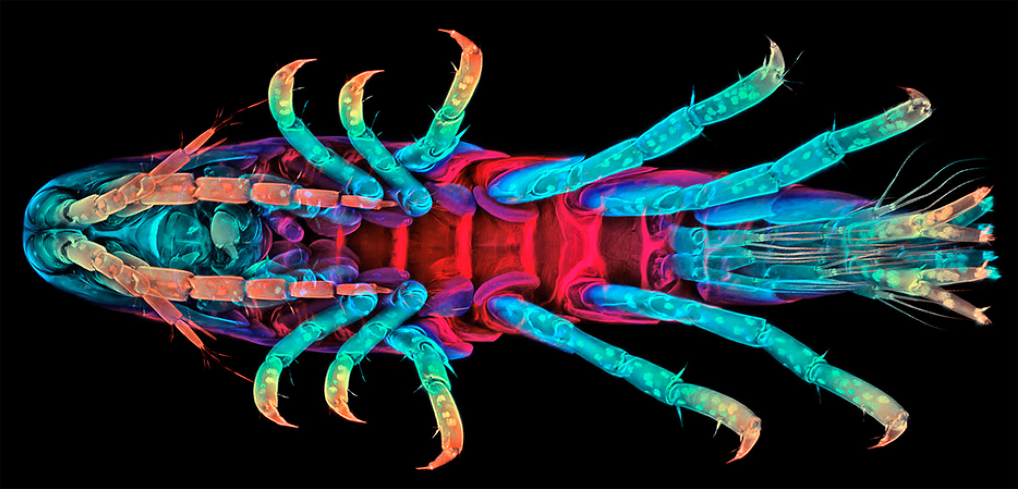
The crustacean amphipod Parhyale hawaiensis has emerged as an attractive experimental model to address longstanding questions in biology, including cell fate specification, segmentation, neurogenesis, organ morphogenesis and regeneration. This site provides comprehensive and easy access to all resources pertaining to Parhyale experimentation.

Genomic resources

Parhyale genome
Contigs were assembled with Abyss at k-length of 70 and 120. Both K70 and K120 assemblies were merged with GAM-NGS. Scaffolding was done with SSPACE. Go here for an ipython notebook of the assembly process.

Scaffolds (.fasta)
Contigs (.fasta)
Annotations (.gff)

Parhyale transcriptome
Gene annotations were done with a combination of Evidence Modeler (EVM) and Augustus. EVM was first used to generate high-confidence predictions using ab initio predictions and transcriptome mapping. Augustus was then trained using these high confidence predictions. Read the first response to this post at BioStars for more detail. Go here for an ipython notebook of the annotation process.

Each gene has an ID in the format of "phaw_30_source_mapped_id". "Source" can either be from transcriptome (tra) or from Augustus gene predictions (aug). "Mapped" can either be unmapped (u) or mapped (m). For example: "phaw_30_tra_u.026676" means this gene is sourced from the transcriptome and does not map to the genome (due to incompleteness of the genome or transcriptome assembly error).

Genes - nucleotide (.fasta)
Genes - protein (.fasta)

Experimental resources

Introduction to Parhyale and animal culture

  • Rehm, E. J., Hannibal, R. L., Chaw, R. C., Vargas-Vila, M. A., & Patel, N. H. (2009). The Crustacean Parhyale hawaiensis: A New Model for Arthropod Development. Cold Spring Harbor Protocols, 2009(1), pdb.emo114–pdb.emo114. http://doi.org/10.1101/pdb.emo114
  • Stamataki, E., & Pavlopoulos, A. (2016). Non-insect crustacean models in developmental genetics including an encomium to Parhyale hawaiensis. Current Opinion in Genetics & Development, 39, 149–156. http://doi.org/10.1016/j.gde.2016.07.004
  • Parhyale hawaiensis culture protocol from Extavour lab

Parhyale embryogenesis and early cell lineage

  • Browne, W. E., Price, A. L., Gerberding, M., & Patel, N. H. (2005). Stages of embryonic development in the amphipod crustacean,Parhyale hawaiensis. Genesis, 42(3), 124–149. http://doi.org/10.1002/gene.20145
  • Gerberding, M., Browne W. E., & Patel N. H. (2002). Cell lineage analysis of the amphipod crustacean Parhyale hawaiensis reveals an early restriction of cell fates. Development, 129(24), 5789–5801. http://doi.org/10.1242/dev.00155
  • Price, A. L., Modrell, M. S., Hannibal, R. L., & Patel, N. H. (2010). Mesoderm and ectoderm lineages in the crustacean Parhyale hawaiensis display intra-germ layer compensation. Developmental Biology, 341(1), 256–266. http://doi.org/10.1016/j.ydbio.2009.12.006
  • Alwes, F., Hinchen, B., & Extavour, C. G. (2011). Patterns of cell lineage, movement, and migration from germ layer specification to gastrulation in the amphipod crustacean Parhyale hawaiensis. Developmental Biology, 359(1), 110–123. http://doi.org/10.1016/j.ydbio.2011.07.029

Dissection, fixation, antibody staining and hybridization of Parhyale embryos

  • Rehm, E. J., Hannibal, R. L., Chaw, R. C., Vargas-Vila, M. A., & Patel, N. H. (2009). Fixation and Dissection of Parhyale hawaiensis Embryos. Cold Spring Harbor Protocols, 2009(1), pdb.prot5127–pdb.prot5127. http://doi.org/10.1101/pdb.prot5127
  • Rehm, E. J., Hannibal, R. L., Chaw, R. C., Vargas-Vila, M. A., & Patel, N. H. (2009). In Situ Hybridization of Labeled RNA Probes to Fixed Parhyale hawaiensis Embryos. Cold Spring Harbor Protocols, 2009(1), pdb.prot5130–pdb.prot5130. http://doi.org/10.1101/pdb.prot5130
  • Rehm, E. J., Hannibal, R. L., Chaw, R. C., Vargas-Vila, M. A., & Patel, N. H. (2009). Antibody Staining of Parhyale hawaiensis Embryos. Cold Spring Harbor Protocols, 2009(1), pdb.prot5129–pdb.prot5129. http://doi.org/10.1101/pdb.prot5129
  • Modified Parhyale in situ hybridization protocol from Averof & Pavlopoulos

Parhyale embryo microinjection and transgenesis

  • Kontarakis, Z., & Pavlopoulos, A. (2014). Transgenesis in Non-model Organisms: The Case of Parhyale. In V. Orgogozo & M. V. Rockman (Eds.), Molecular Methods for Evolutionary Genetics (Vol. 1196, pp. 145–181). New York, NY: Springer New York. http://doi.org/10.1007/978-1-4939-1242-1_10
  • Pavlopoulos, A., & Averof, M. (2005). Establishing genetic transformation for comparative developmental studies in the crustacean Parhyale hawaiensis. Proceedings of the National Academy of Sciences of the United States of America, 102(22), 7888–7893. http://doi.org/10.1073/pnas.0501101102
  • Pavlopoulos, A., Kontarakis, Z., Liubicich, D. M., Serano, J. M., Akam, M., Patel, N. H., & Averof, M. (2009). Probing the evolution of appendage specialization by Hox gene misexpression in an emerging model crustacean. Proceedings of the National Academy of Sciences of the United States of America, 106(33), 13897–13902. http://doi.org/10.1073/pnas.0902804106
  • Kontarakis, Z., Pavlopoulos, A., Kiupakis, A., Konstantinides, N., Douris, V., & Averof, M. (2011). A versatile strategy for gene trapping and trap conversion in emerging model organisms. Development, 138(12), 2625–2630. http://doi.org/10.1242/dev.066324
  • Rehm, E. J., Hannibal, R. L., Chaw, R. C., Vargas-Vila, M. A., & Patel, N. H. (2009). Injection of Parhyale hawaiensis Blastomeres with Fluorescently Labeled Tracers. Cold Spring Harbor Protocols, 2009(1), pdb.prot5128–pdb.prot5128. http://doi.org/10.1101/pdb.prot5128

Parhyale blastomere ablation and isolation

  • Extavour, C. G. (2005). The fate of isolated blastomeres with respect to germ cell formation in the amphipod crustacean Parhyale hawaiensis. Developmental Biology, 277(2), 387–402. http://doi.org/10.1016/j.ydbio.2004.09.030
  • Alwes, F., Hinchen, B., & Extavour, C. G. (2011). Patterns of cell lineage, movement, and migration from germ layer specification to gastrulation in the amphipod crustacean Parhyale hawaiensis. Developmental Biology, 359(1), 110–123. http://doi.org/10.1016/j.ydbio.2011.07.029
  • Hannibal, R. L., Price, A. L., & Patel, N. H. (2012). The functional relationship between ectodermal and mesodermal segmentation in the crustacean, Parhyale hawaiensis. Developmental Biology, 361(2), 427–438. http://doi.org/10.1016/j.ydbio.2011.09.033
  • Nast, A. R., & Extavour, C. G. (2014). Ablation of a Single Cell From Eight-cell Embryos of the Amphipod Crustacean arhyale hawaiensis. Journal of Visualized Experiments, (85), 1–8. http://doi.org/10.3791/51073

CRISPR/Cas genome editing in Parhyale embryos

  • Kao, D., Lai, A. G., Stamataki, E., Rosic, S., Konstantinides, N., Jarvis, E., et al. (2016). The genome of the crustacean Parhyale hawaiensis, a model for animal development, regeneration, immunity and lignocellulose digestion. eLife, 5, e20062–45. http://doi.org/10.7554/eLife.20062
  • Martin, A., Serano, J. M., Jarvis, E., Bruce, H. S., Wang, J., Ray, S., et al. (2016). CRISPR/Cas9 Mutagenesis Reveals Versatile Roles of Hox Genes in Crustacean Limb Specification and Evolution. Current Biology, 26(1), 14–26. http://doi.org/10.1016/j.cub.2015.11.021
  • Serano, J. M., Martin, A., Liubicich, D. M., Jarvis, E., Bruce, H. S., La, K., et al. (2016). Comprehensive analysis of Hox gene expression in the amphipod crustacean Parhyale hawaiensis. Developmental Biology, 409(1), 297–309. http://doi.org/10.1016/j.ydbio.2015.10.029

Parhyale embryo live imaging

  • Price, A. L., & Patel, N. H. (2007). Investigating divergent mechanisms of mesoderm development in arthropods: the expression ofPh-twist andPh-mef2 inParhyale hawaiensis. Journal of Experimental Zoology Part B: Molecular and Developmental Evolution, 310B(1), 24–40. http://doi.org/10.1002/jez.b.21135
  • Alwes, F., Hinchen, B., & Extavour, C. G. (2011). Patterns of cell lineage, movement, and migration from germ layer specification to gastrulation in the amphipod crustacean Parhyale hawaiensis. Developmental Biology, 359(1), 110–123. http://doi.org/10.1016/j.ydbio.2011.07.029
  • Wolff, C., Tinevez, J.-Y., Pietzsch, T., Stamataki, E., Harich, B., Preibisch, S., et al. (2017). Reconstruction of cell lineages and behaviors underlying arthropod limb outgrowth with multi-view light-sheet imaging and tracking. BioRxiv 1–54. http://doi.org/10.1101/112623

Community

Aziz Aboobaker lab
University of Oxford
Department of Zoology
South Parks Road Oxford, UK OX13PS
Email aziz.aboobaker@zoo.ox.ac.uk
Web page http://aboobakerlab.com

Michalis Averof Lab
Institute of Functional Genomics Lyon (IGFL)
32-34 avenue Tony Garnier
Lyon 69007, France
Tel. +33-4-26731364
Email michalis.averof@ens-lyon.fr
Web page https://averof-lab.org/

Cassandra G. Extavour Lab
Harvard University
Department of Organismic and Evolutionary Biology
Department of Molecular and Cellular Biology
16 Divinity Avenue, BioLabs 2087
Cambridge, MA 02138, USA
Tel. Office 1 617 496 1935
Tel. Lab 1 617 496 0983/2984
Email extavour@oeb.harvard.edu
Web page http://www.extavourlab.com

Nipam H. Patel Lab
University of California, Berkeley
Department of Molecular & Cell Biology
525A LSA #3200
Berkeley, CA 94720-3200, USA
Tel. Office +1-510-643-4605
Tel. Lab +1-510-643-4201
Email nipam@berkeley.edu
Web pagehttp://www.patellab.org

Anastasios Pavlopoulos Lab
Janelia Research Campus
19700 Helix Drive
Ashburn, VA 20147, USA
Tel. +1-571-209-4000 (ext.3337)
Email pavlopoulosa@janelia.hhmi.org
Web page https://www.janelia.org/lab/pavlopoulos-lab

Carsten Wolff / Gerhard Scholtz Lab
Humboldt University
Department of Biology / Comparative Zoology
Philippstr. 13, Haus 2
10115 Berlin, Germany
Tel. +49-30-2093-6284
Email carsten.wolff@rz.hu-berlin.de
Web page https://www.biologie.hu-berlin.de/de/gruppenseiten/compzool